
21st century tumor models

Cancer cell lines need a makeover

Currently, all stages of drug discovery and basic molecular cancer research rely on established cancer cell lines. The most comprehensively utilized collection of established cancer cell lines, composing the CCLE collection and underlying e.g., the DepMap data, consist of around 2000 cancer cell lines. The median year of establishment of these cell lines is 1991, pre-dating development of targeted cancer therapies and spanning all the way back to the 1950s. Many key cellular properties such lineage heterogeneity, genomic stability and mutational burden change during prolonged culture of cancer cell lines and consequently, the discovery and research of novel therapeutic targets and analysis of e.g., drug resistance mechanism can be significantly biased. Novel cancer cell lines and tumor models representing patients treated with the current cancer therapies are therefore urgently needed to allow more clinically relevant research of the efficacy and resistance mechanism to the latest novel cancer therapies.

MISB Cancer Cell Line Panel

21st century cancer research requires 21st century model cell lines

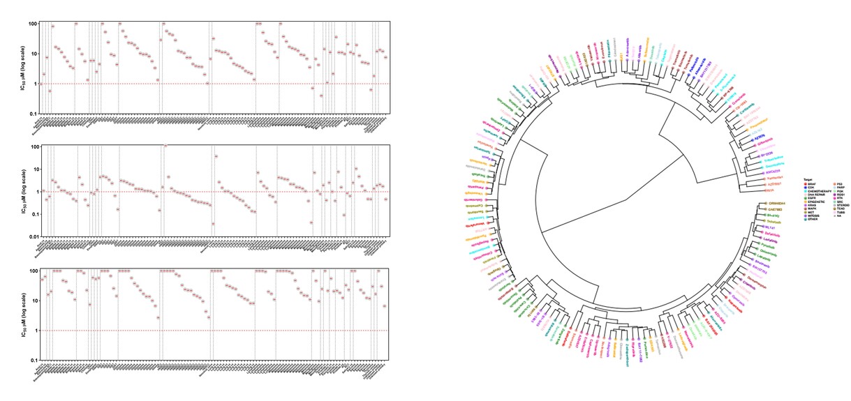
In context of our ongoing clinical research activities, we are building a comprehensive collection of new cancer model cell lines to serve the needs of cancer research and drug development. In parallel to collection of tumor samples for ex vivo drug efficacy testing, we´ve established a proprietary cell line repository consisting of 150+ cell lines representing 50+ different solid cancer typess (@01/2026); the MISB Cancer Cell Line Panel. The continuously expanding MISB cell line panel has been characterized extensively in context of Misvik’s Precision Oncology initiative including using DNA and RNA sequencing and extensive drug screens of hundreds of cancer therapies, including the very latest novel cancer drugs. The MISB cell line panel offers a collection of low-passage clinically relevant tumor cell models that retain the original patient-specific tumor biology including lineage heterogeneity, genetic background and treatment history (62 different therapies covered as previous line of treatment before relapse and establishment of cell line). The MISB cell line panel is offered for rapid large scale high-throughput cell panel screening, in-depth studies of drug efficacy and combinatorial synergy as well as for genome-wide loss of function genomic screening using siRNA and CRISPR.


Common and rare cancers

The common cancer types included in the current MISB cell line panel (@01/2026) include: Bladder, Breast, Cholangiocarcinoma, Colon, Esophageal, Endometrial, Glioma, Head and Neck, Lung, Melanoma, Ovarian, Pancreatic, Prostate and Renal. Rare cancer types include: Conjunctival melanoma, Choroidal melanoma, DSRCT, GIST, Ileocecal valve, Large Cell Neuroendocrine PCa, Salivary gland, Sarcomas, Urachal and cancers of unknown origin. 


Pan-cancer including first from indication

Diverse set of common and rare solid cancers covering 50+ different cancer types


Extensive characterization

Detailed molecular and clinical profiling including DNA & RNAseq, proteomics and large-scale drug sensitivity profile


Targeted therapy relevance

Established from tumors of heavily pretreated patients covering 64 targeted therapies


Proprietary

No license fees = competitive service pricing and freedom to operate


Patient derived ex vivo tumor models

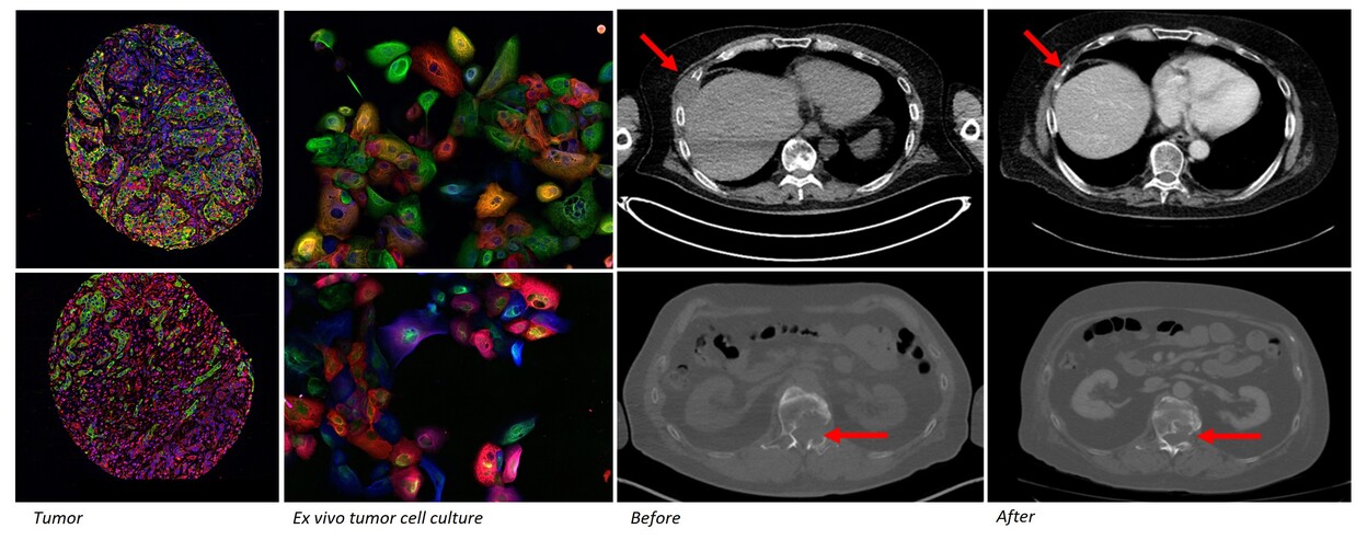
Patient derived functional tumor models retain the cellular composition and differentiation state heterogeneity of the parental tumors. Primary ex vivo models also reflect the  pathology and clinical behavior of the cancer. Misvik´s ex vivo tumor models are a continuously expanding collection of early passage patient derived functional tumor models cryopreserved following initial tissue dissociation and establishment of organoid culture. The ex vivo model collection currently contains 700+ tumor samples representing 90+ different solid cancer types including rare to ultra rare cancers. The sample series also contains matched longitudinal samples from the same patient prior and after treatment allowing study of therapy efficacy in treatment naive and relapsed tumor models. All samples are incorporated on Misvik´s master RPPA (reverse-phase-protein array) sample repository for rapid target evaluation and general protein expression analysis across different cancers types.

Comprehensively annotated and fully characterized

Ex vivo tumor models reflect disease characteristics of the current patient populations receiving latest novel cancer therapies as the standard care. All available clinical annotations are collected from all ex vivo models and coupled with next-generation molecular profiling and histopathology. This allows selection and study of models that recapitulate the research question and clinical need of today´s oncology drug discovery and cancer research. The ex vivo drug screening results are always correlated with the available clinical, molecular and histopathological information to facilitate biomarker discovery and indication categorization. The ex vivo tumor models are available for large-scale pan-cancer screens or standalone studies of any molecular entity including novel immuno-oncology therapies as the models incorporate the tumor tissues full cellular complexity.

Building insights on latest novel cancer drugs using pan-cancer ex vivo tumor models.

Biologically relevant ex vivo tumor models are primary cultures isolated directly from dissociated tumor samples

References

  1. Pan-cancer ex vivo target evaluation of phosphodiesterase 3 A (PDE3A). Rice K, Lehtinen, N., Välimäki, E. et al. J Mol Med 2026;104(69). https://doi.org/10.1007/s00109-026-02677-7
  2. FANCD2 restrains fork progression and prevents fragility at early origins upon re-replication. Badra-Fajardo N, Karydi E, Bayona-Feliu A, Gómez-González B, Preza O, Arbi M, Kalogeropoulou A, Rantala JK, Taraviras S, Aguilera A, Lygerou Z. Nat Commun. 2026;17(1):2478. doi: 10.1038/s41467-026-68966-4.
  3. LIMA1-alpha staining predicts curative intent surgery response in HPV negative head and neck cancer.Qiao X, Routila J, Tienhaara M, Irjala H, Santhi PP, Huusko T, Nissi L, Paatero I, Lehtinen N, Rantala J, Viljanen T, Leivo I, Koivunen P, Jouppila-Mättö A, Taulu R, Bäck L, Wilkman T, Haapio E, Kinnunen I, Kurppa K, Westermarck J, Ventelä S. EMBO Mol Med. 2025;(8):2095-2114. doi: 10.1038/s44321-025-00266-8.
  4. In-vitro assays for immuno-oncology drug efficacy assessment and screening for personalized cancer therapy: scopes and challenges. Rahman MM, Wells G, Rantala JK, Helleday T, Muthana M, Danson SJ. Expert Rev Clin Immunol. 2024;(8):821-838. doi: 10.1080/1744666X.2024.2336583.
  5. Ex-vivo drug screening of surgically resected glioma stem cells to replace murine avatars and provide personalise cancer therapy for glioblastoma patients. Gagg H, Williams ST, Conroy S, Myers KN, McGarrity-Cottrell C, Jones C, Helleday T, Rantala J, Rominiyi O, Danson SJ, Collis SJ, Wells G. F1000Res. 2024;12:954. doi: 10.12688/f1000research.
  6. Assessment of targeted therapy opportunities in sinonasal cancers using patient-derived functional tumor models. Lehtinen N, Suhonen J, Rice K, Välimäki E, Toriseva M, Routila J, Halme P, Rahi M, Irjala H, Leivo I, Kallajoki M, Nees M, Kuopio T, Ventelä S, Rantala JK. Transl Oncol. 2024;44:101935.
  7. Defined extracellular matrix compositions support stiffness-insensitive cell spreading and adhesion signaling. Conway JRW, Isomursu A, Follain G, Härmä V, Jou-Ollé E, Pasquier N, Välimäki EPO, Rantala JK, Ivaska J. Proc Natl Acad Sci U S A. 2023;120(43):e2304288120.
  8. FBXL12 degrades FANCD2 to regulate replication recovery and promote cancer cell survival under conditions of replication stress Brunner A, Li Q, Fisicaro S, Kourtesakis A, Viiliäinen J, Johansson HJ, Pandey V, Mayank AK, Lehtiö J, Wohlschlegel JA, Spruck C, Rantala JK, Orre LM, Sangfelt O.. Mol Cell. 2023 Oct 19;83(20):3720-3739.e8.
  9. Precision oncology using ex vivo technology: a step towards individualised cancer care? Williams ST, Wells G, Conroy S, Gagg H, Allen R, Rominiyi O, Helleday T, Hullock K, Pennington CEW, Rantala J, Collis SJ, Danson SJ. Expert Rev Mol Med. 2022;3:1-48.
  10. Cisplatin overcomes radiotherapy resistance in OCT4-expressing head and neck squamous cell carcinoma. Routila J, Qiao X, Weltner J, Rantala JK, Carpén T, Hagström J, Mäkitie A, Leivo I, Ruuskanen M, Söderlund J, Rintala M, Hietanen S, Irjala H, Minn H, Westermarck J, Ventelä S. Oral Oncol. 2022;127:105772.
  11. FBXO7/EYA2-SCFFBXW7 axis promotes AXL-mediated maintenance of mesenchymal and immune evasion phenotypes of cancer cells. Shen JZ, Qiu Z, Wu Q, Zhang G, Harris R, Sun D, Rantala J, Barshop WD, Zhao L, Lv D, Won KA, Wohlschlegel J, Sangfelt O, Laman H, Rich JN, Spruck C. A Mol Cell. 2022;82(6):1123-1139.e8.
  12. Ex Vivo Drug Screening Informed Targeted Therapy for Metastatic Parotid Squamous Cell Carcinoma. Nykänen N, Mäkelä R, Arjonen A, Härmä V, Lewandowski L, Snowden E, Blaesius R, Jantunen I, Kuopio T, Kononen J, Rantala JK. Front Oncol. 2021;11:735820.
  13. FBXO44 promotes DNA replication-coupled repetitive element silencing in cancer cells. Shen JZ, Qiu Z, Wu Q, Finlay D, Garcia G, Sun D, Rantala J et al. Cell. 2021;184(2):352-369.e23.
  14. Ex vivo analysis of DNA repair targeting in extreme rare cutaneous apocrine sweat gland carcinoma. Mäkelä R, Härmä V, Badra Fajardo N, Wells G, Lygerou Z, Sangfelt O, Kononen J, Rantala JK. Oncotarget. 2021;12(11):1100-1109.
  15. Substrate-biased activity-based probes identify proteases that cleave receptor CDCP1Kryza T, Khan T, Lovell S, Harrington BS, Yin J, Porazinski S, Pajic M, Koistinen H, Rantala JK et al.. Nat Chem Biol. 2021;Apr 15.
  16. Ex vivo assessment of targeted therapies in a rare metastatic epithelial-myoepithelial carcinoma. Mäkelä R, Arjonen A, Suryo Rahmanto A, Härmä V, Lehtiö J, Kuopio T, Helleday T, Sangfelt O, Kononen J, Rantala JK. Neoplasia. 2020;22(9):390-398.
  17. PTEN and DNA-PK determine sensitivity and recovery in response to WEE1 inhibition in human breast cancer. Brunner A, Suryo Rahmanto A, Johansson H, Franco M, Viiliäinen J, Gazi M, Frings O, Fredlund E, Spruck C, Lehtiö J, Rantala JK, Larsson LG, Sangfelt O. Elife. 2020;9:e57894.
  18. Ex vivo modelling of therapy efficacy for rare Krugenberg tumors – a report of two cases. Arjonen A, Mäkelä R, Virtakoivu R, Härmä V, Kuopio T, Hakkarainen H, Hollmén M, Kononen J, Rantala JK. Clin. Onc. Res. 2020;3(7).
  19. Ex vivo modelling of drug efficacy in a rare metastatic urachal carcinoma. Mäkelä R, Arjonen A, Härmä V, Rintanen N, Paasonen L, Paprotka T, Rönsch K, Kuopio T, Kononen J, Rantala JK. BMC Cancer. 2020;20(1):590.
  20. Image-based ex vivo drug screen to assess targeted therapies in recurrent thymoma. Arjonen A, Mäkelä R, Härmä V, Rintanen N, Kuopio T, Kononen J, Rantala JK. Lung Cancer. 2020;145:27-32.
  21. Clonal Evolution of MEK/MAPK Pathway Activating Mutations in a Metastatic Colorectal Cancer CaseLehtomaki KI, Lahtinen LI, Rintanen N, Kuopio T, Kholova I, Mäkelä R, Rantala JK, Kellokumpu-Lehtinen PL, Kononen J. Anticancer Res. 39(11):5867-5877. 2019.
  22. Personalized Drug Sensitivity Screening for Bladder Cancer Using Conditionally Reprogrammed Patient-derived Cells. Kettunen K, Boström PJ, Lamminen T, Heinosalo T, West G, Saarinen I, Kaipio K, Rantala JK, Albanese C Poutanen M, Taimen P. Eur. J. Urol. 76(4):430-434. 2019.
  23. Combined prognostic value of CD274 (PD-L1)/PDCDI (PD-1) expression and immune cell infiltration in colorectal cancer as per mismatch repair status. Ahtiainen M, Wirta EV, Kuopio T, Seppälä T, Rantala JK, Mecklin JP, Böhm J. Mod Pathol. 32(6):866-883. 2019.
  24. Rantala JK. Diagnostic therapy efficacy assessment in genomic medicine and rare cancer care. Sci. Tech. 3(28). 2018.